Histology Manager
Queensland Brain Institute
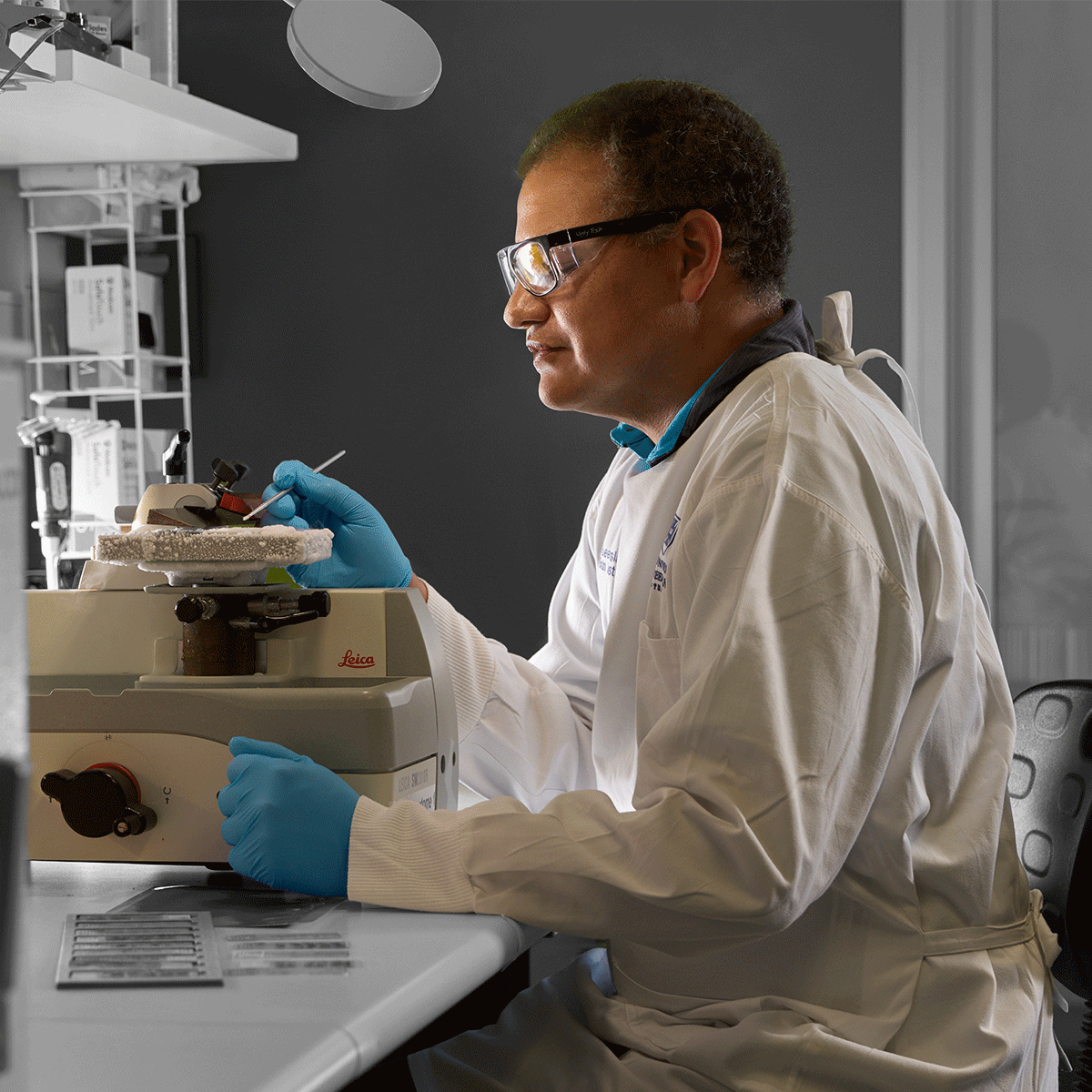
"I am the histology manager for the Queensland Brain Institute. I have spent three decades in neorscience research and training the future experts in research histology."
"Creativity is the core of how I solve problems in research and how I approach teaching and learning. Without creativity, nothing new would be dreamt into reality."
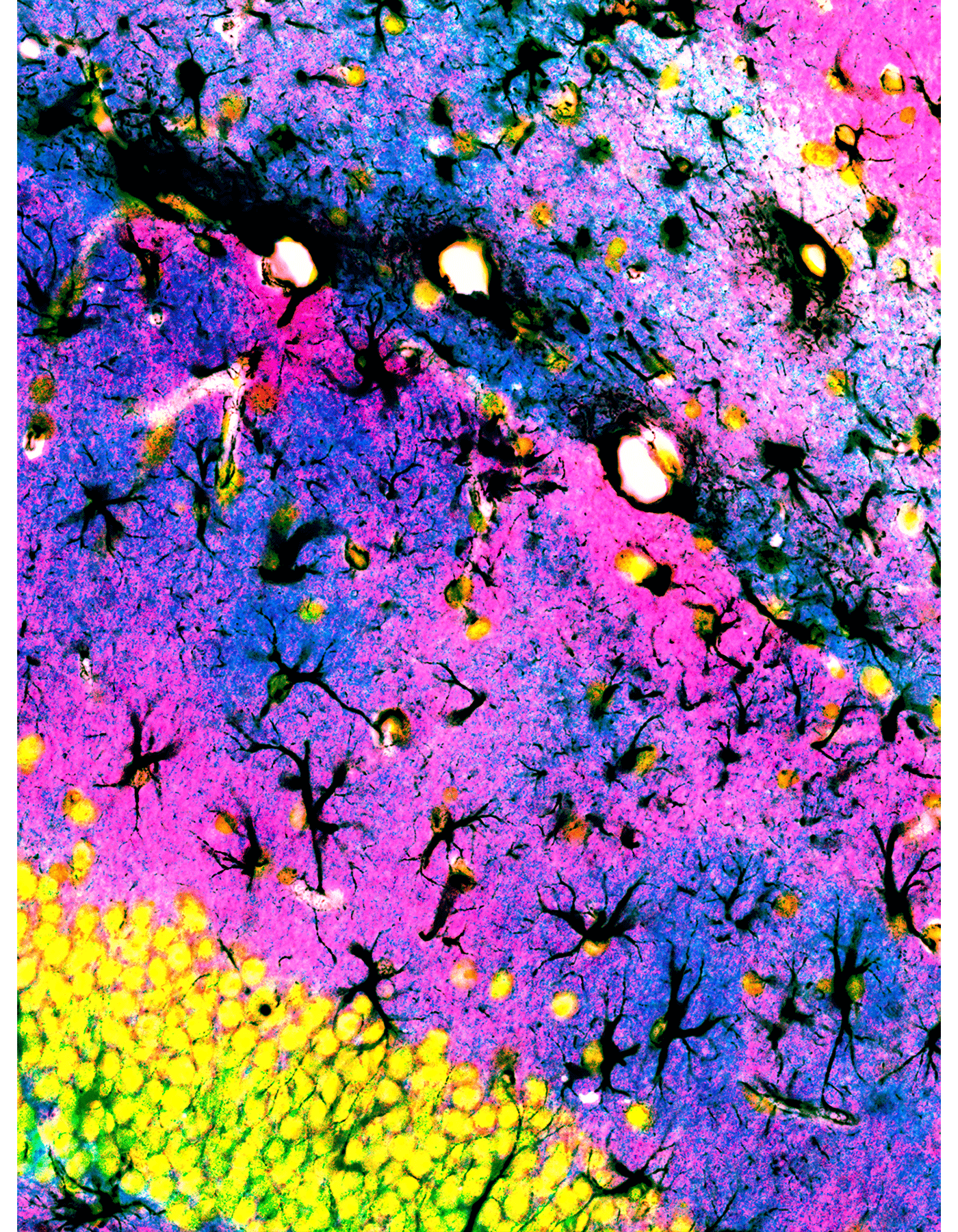
Black branches and trunks of astrocytes holding up pink and blue islands of membrane transporters that regulate excitatory synaptic transmission of the yellow neurons.
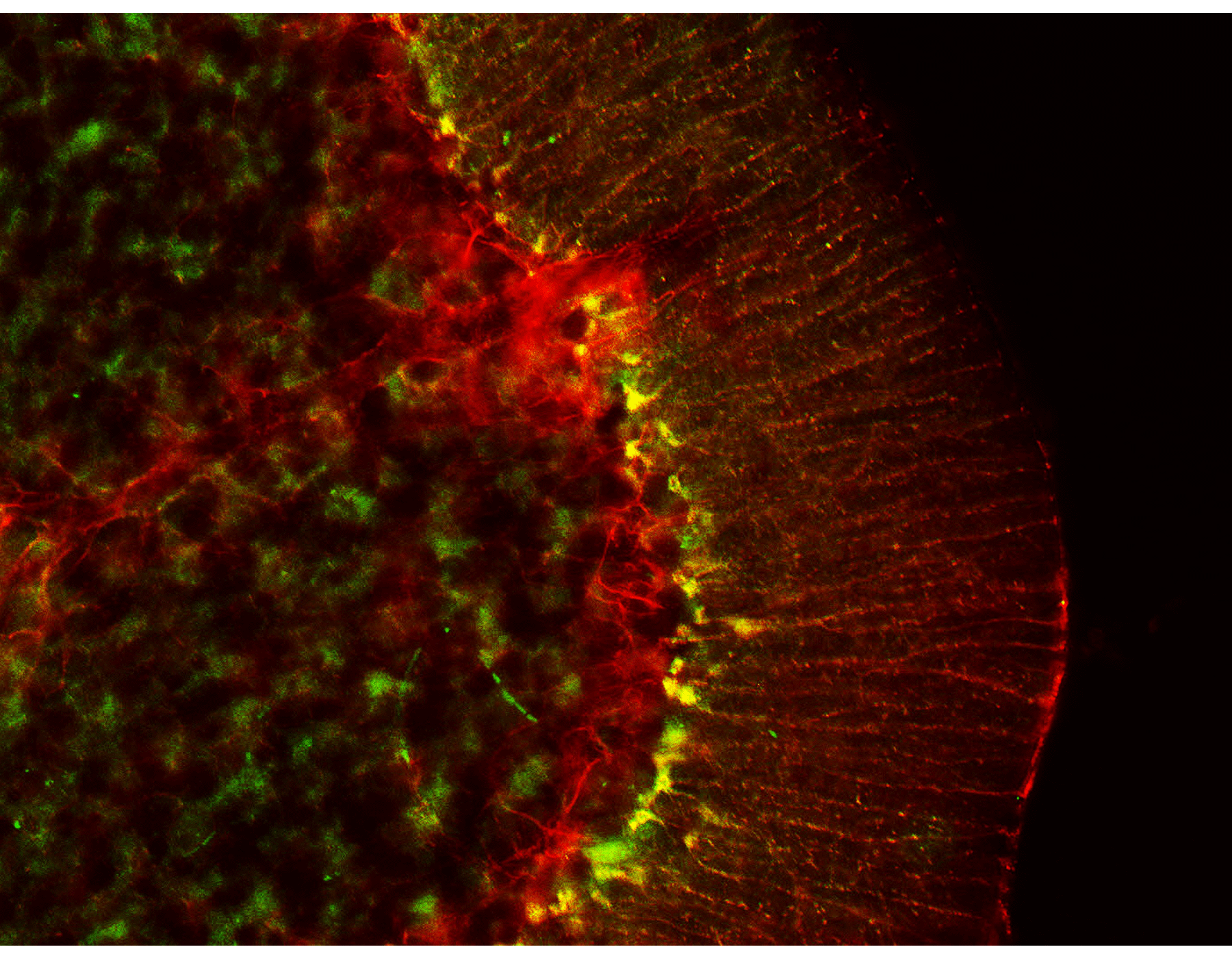
Glial cells have numerous shapes, which provide insights into their various functional roles in thebrain. Bergmann glia (orange) have radial processes for guidance and structure, while star-shaped astrocytes (green and red) wrap blood vessels and neurons.
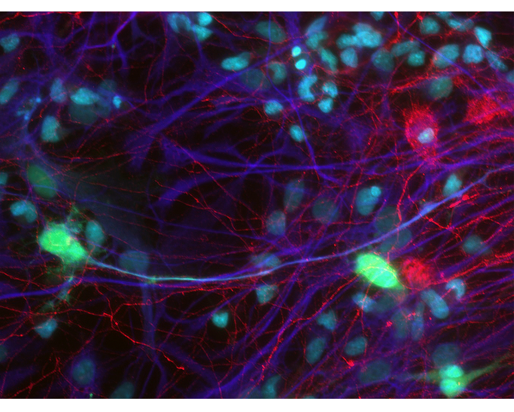
Cortical brain cultures are commonly used to understand how neurons and glia cells communicate. Glia processes (dark blue) make a web that bridge together islands of neurons with pale blue nuclei and a network of interconnecting fibers (red).
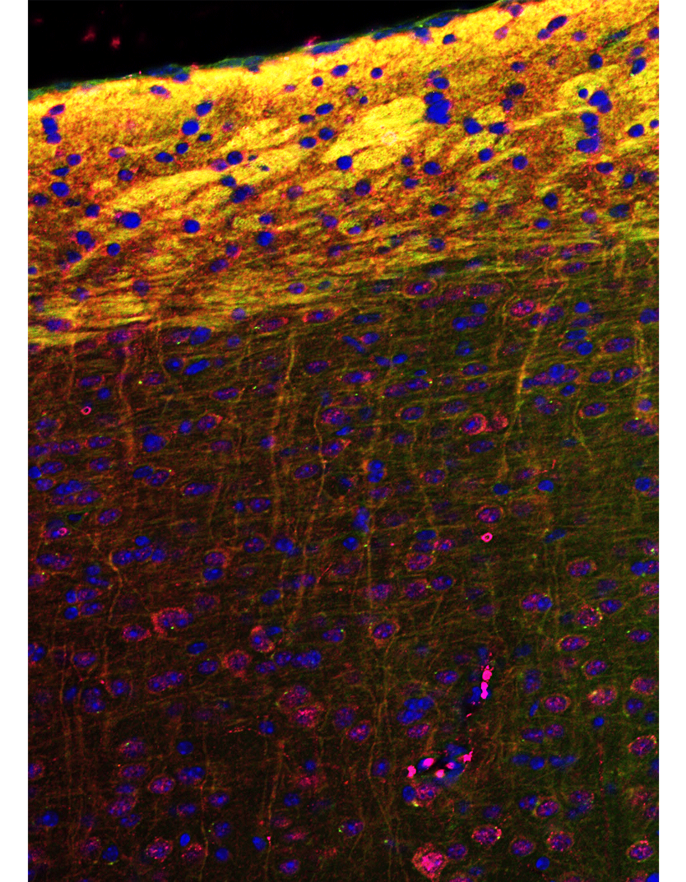
The cells (blue) in the brain communicate through axons that share common pathways that look like thunder clouds (yellow). These axons project long distances to make electrical synapse that connected distal regions of the body.
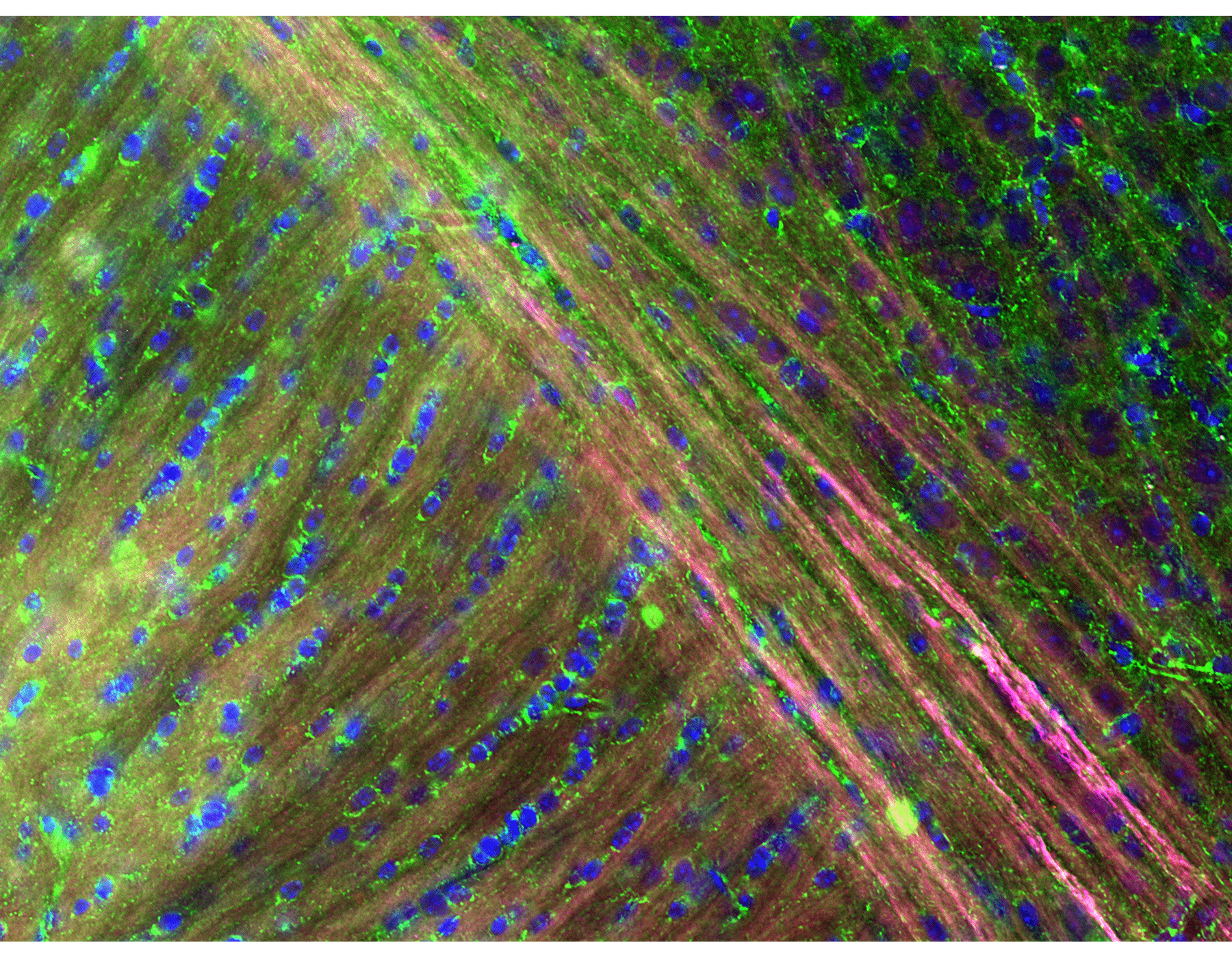
Information in the brain travels through numerous processes (magenta) in all different directions that merge together into a super highway.
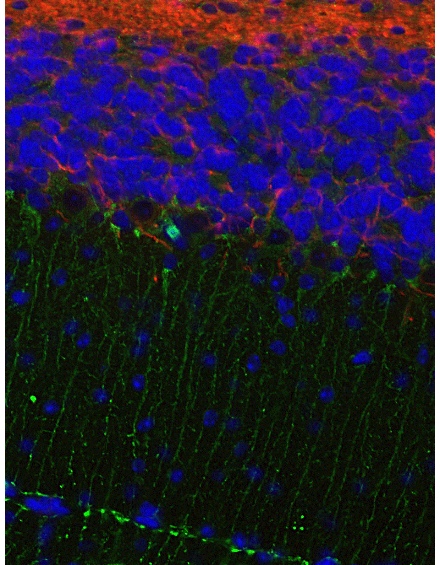
Movement is regulated by some of the smallest and largest neurons (blue) in the cerebellum, which is highly organised into distinct layers. The radial glia (green) and the astrocytes (red) have a critical supportive role and define the different layers.
"When taking images for research I try to limit the images to regions that answer one question. However, when investigating the complex nature of a brain section it's very hard to keep an image simple. Highlighting the regular distributions of glial cells provides order to clouds of membranes that surround the neighbouring invisible neurons."