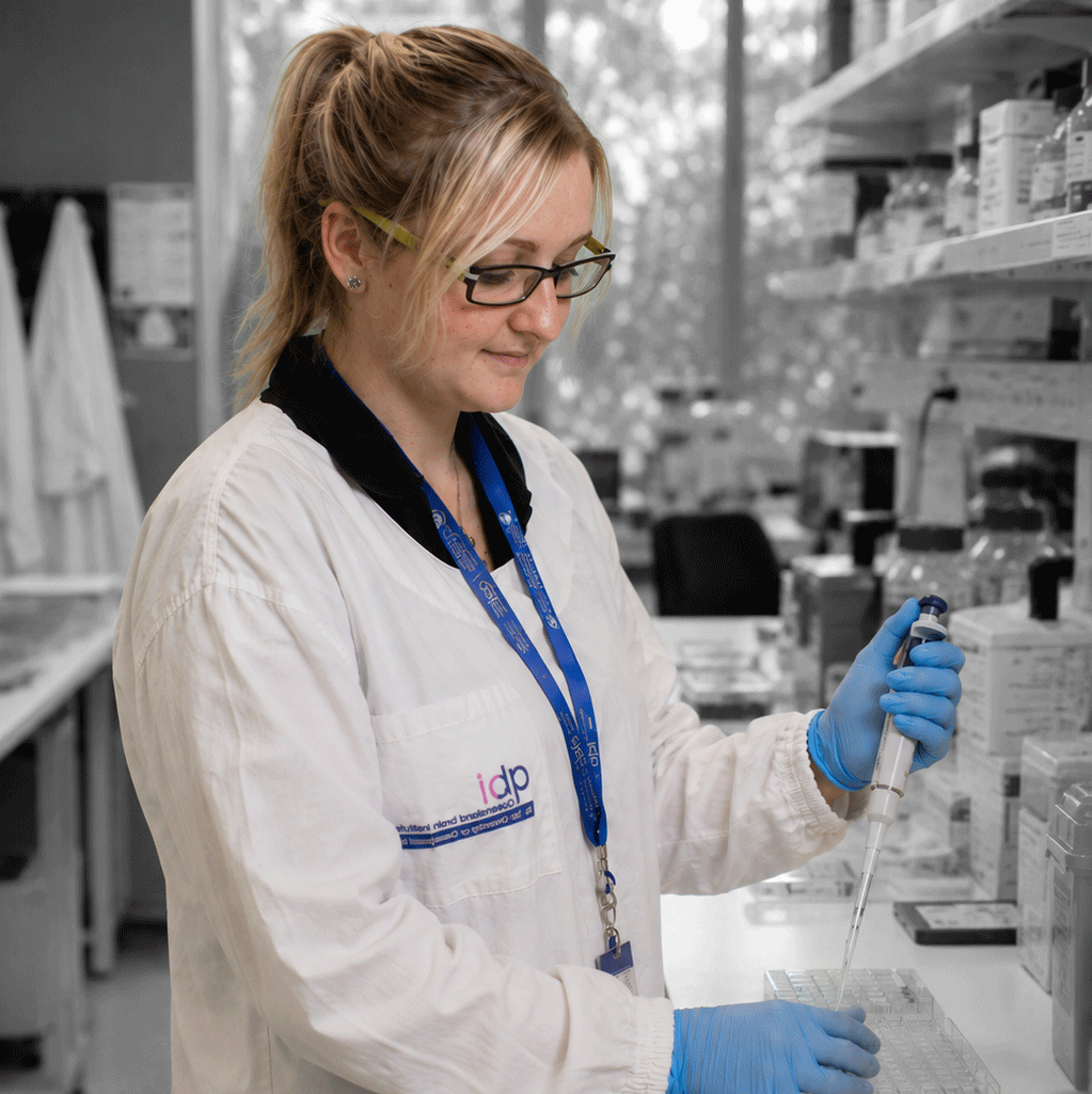
Dr Rachel Gormal
Senior Research Officer
Meunier lab
Queensland Brain Institute
"Scientific research is driven not only by rigorous investigation but also by imagination and creative thinking."
Senior Research Officer
Meunier lab
Queensland Brain Institute
"Scientific research is driven not only by rigorous investigation but also by imagination and creative thinking."
"My creative pursuits outside the laboratory continually influence the way I think within it. Experiences beyond neuroscience encourage new perspectives, remind me to remain curious, and often shape the questions I ask."
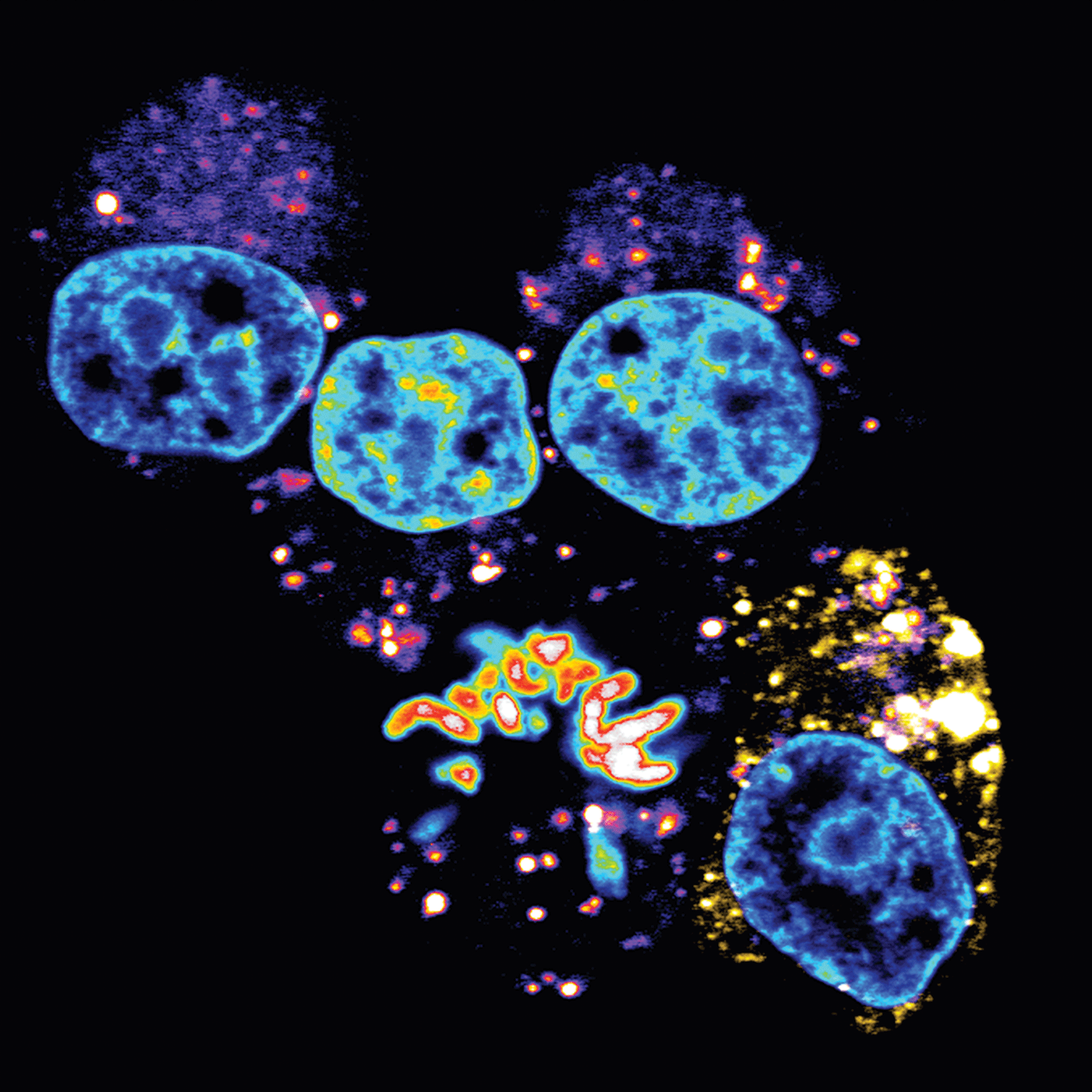
This microscope image shows five individual cells. Four of the cells contain a moon-like blue structure each, which is the cell nucleus, the control centre of the cell, where its DNA is stored as chromosomes. In one cell, the nucleus is beginning to break down, marking the start of cell division. This brief stage allows the chromosomes to separate so they can replicate before the cell divides in two. The image was pseudocoloured to show dense areas in different shades.
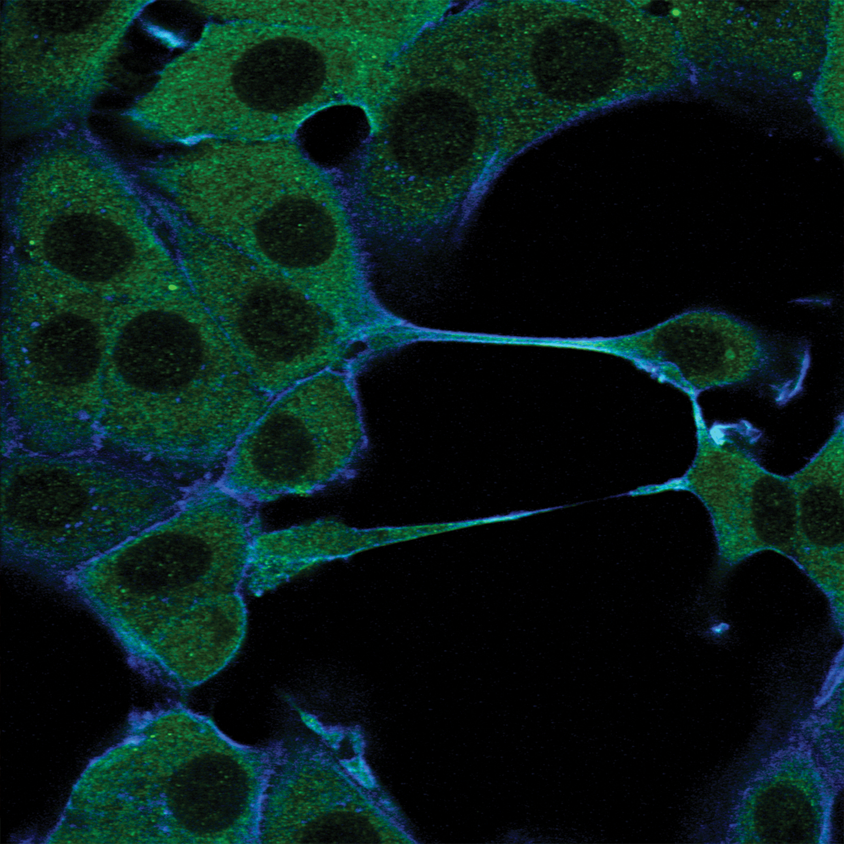
This microscope image shows cultured cells growing in a laboratory dish. Two proteins have been labelled using fluorescent antibodies to reveal their location within the cells. The protein shown in green is distributed throughout the cell, but is absent from the nucleus, the cell's control centre. This creates the appearance of dark openings, or "holes," within each cell. A second protein, shown in blue, is found only along the cell's outer boundary, outlining its shape and defining the cell's edge.
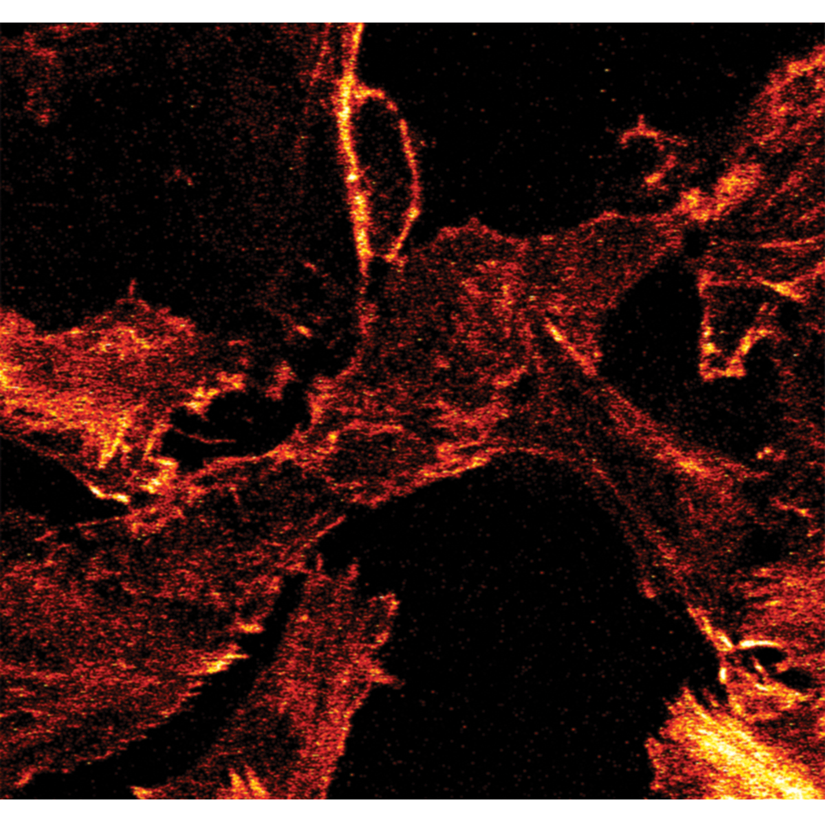
This microscope image highlights actin fibres within cultured cells, shown in shades of red and orange. Actin is a structural protein that plays a vital role in many cellular processes, helping cells maintain their shape while also supporting movement and communication. The brightest regions reveal areas whereactin is densely concentrated, around the cell's outer edge or within thick bundles that span the cell. As these actin filaments assemble into long, tightly packed structures, they create striking pointed formations that resemble glowing, red-hot flames.
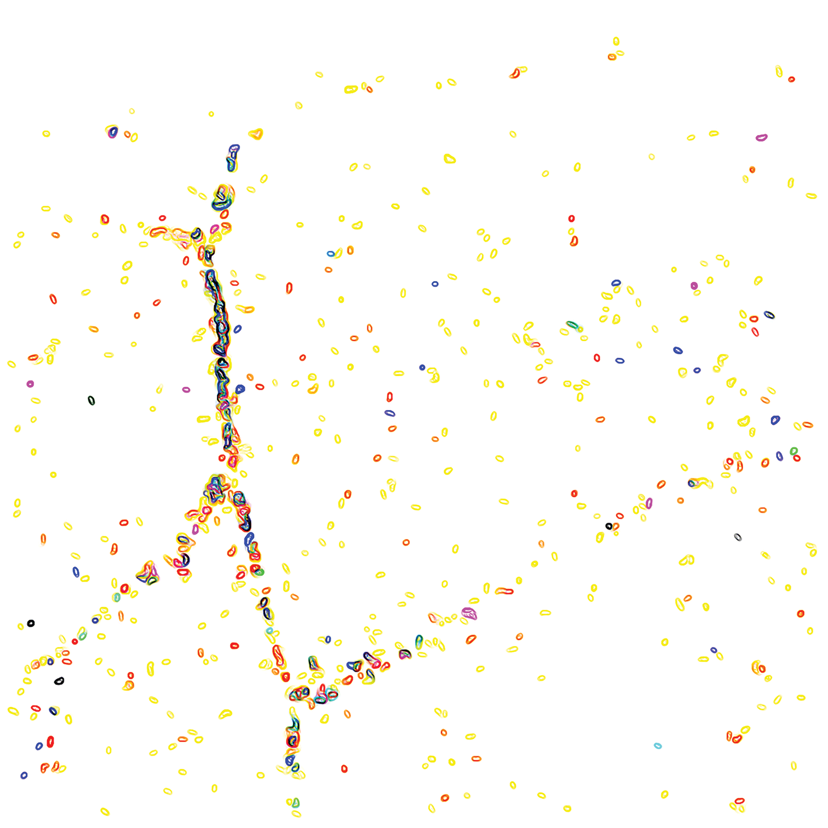
Localising the connection between cells in a monolayer by E-cadherin. This image combines CRISPR genetic engineering and super-resolution technology.
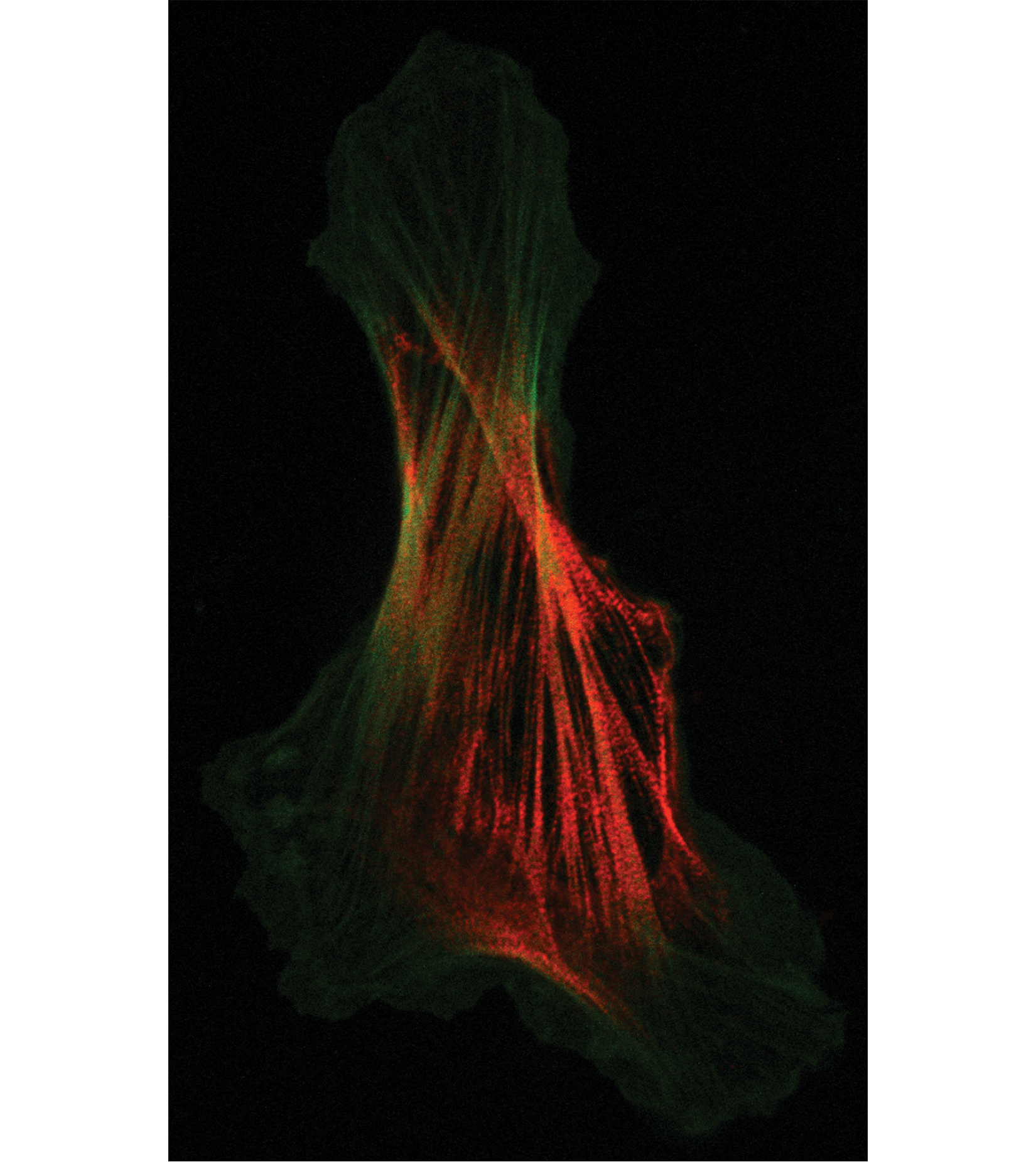
The image symbolises the relationship between science and art; just as our creativity emerges from billions of interconnected brain cells, scientific discovery begins with imagination, and discoveries are built by connecting countless small insights into transformative breakthroughs. It reminds us that the scientific process is not only rigorous and analytical, but also deeply imaginative, celebrating the creativity that lies at the heart of both science and the human experience.
"Scientific research is driven not only by rigorous investigation, but by imagination and creative thinking. In neuroscience, microscopy reveals structures beyondthe limits of human vision, offering a glimpse into the intricate beauty that exists at the microscopic scale. These images explore the architecture of cells, where form and function are intimately connected."
Dr. Rachel Gormal obtained a Bachelor of Science Majoring in Neuroscience with Honours in 2009 from the University of Queensland. She continued her work in the Single Molecule Neuroscience Laboratory led by Professor Frederic Meunier, working as a Research Assistant and then Lab manager for over a decade before obtaining her PhD at Griffith University in 2022. Her research focus is primarily on the study of neuro endo- and exo-cytosis mechanisms, by applying molecular biology and super-resolution microscopy approaches.




.png)