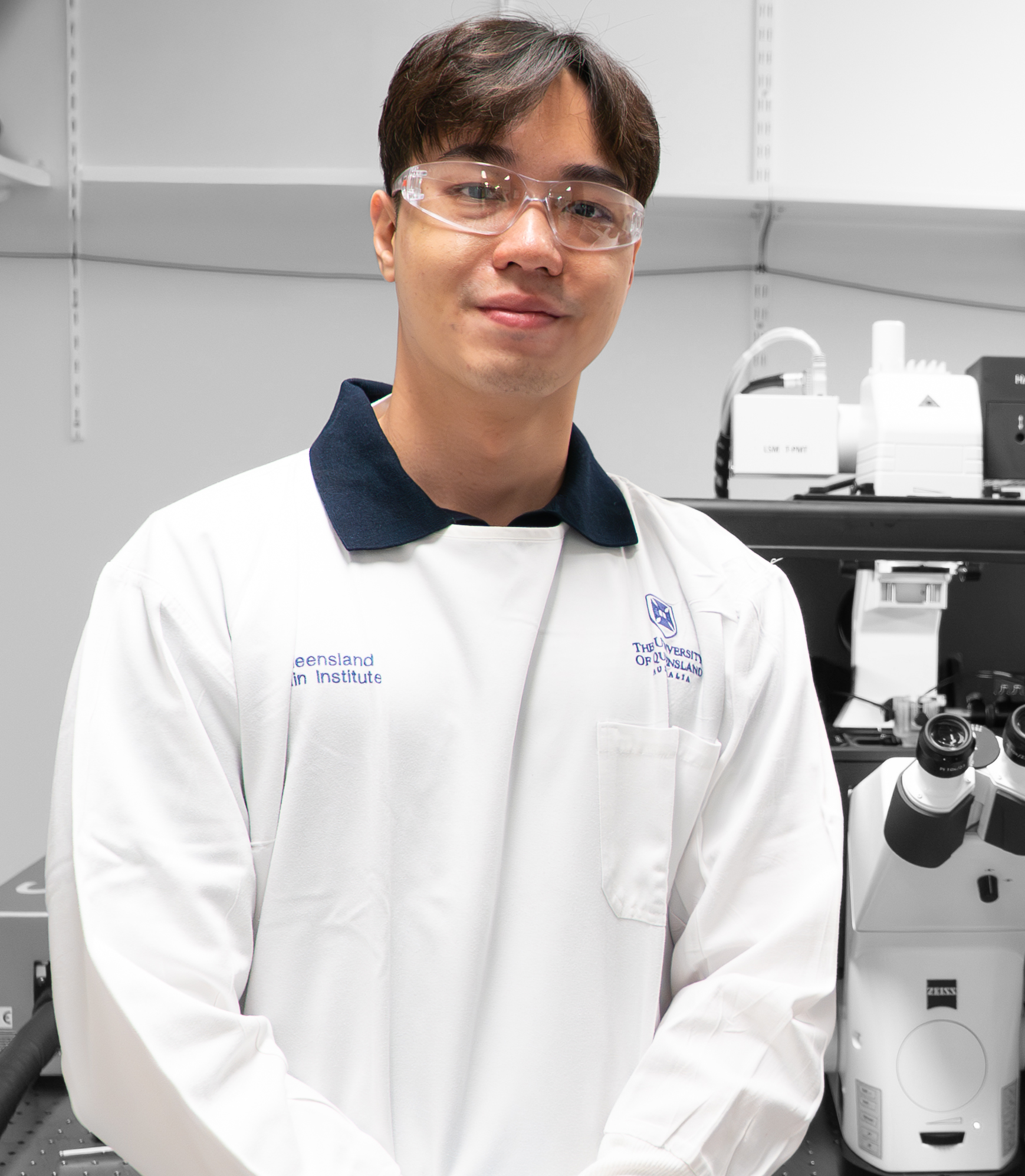
Dr Jonathan Lee
Postdoctoral researcher
Padmanabhan lab
Queensland Brain Institute
"I am a postdoctoral research fellow in the Molecular and Systems Medicine Group at the School of Biomedical Sciences and Queensland Brain Institute."
Postdoctoral researcher
Padmanabhan lab
Queensland Brain Institute
"I am a postdoctoral research fellow in the Molecular and Systems Medicine Group at the School of Biomedical Sciences and Queensland Brain Institute."
"Science constantly requires us to innovate, problem-solve and test unconventional ideas, all of which stem from curiosity and creativity. My creativity in science is often inspired by spotting new, interesting patterns within cells in the diseased state, which motivates me to probe further to understand how these proteins become dysfunctional."
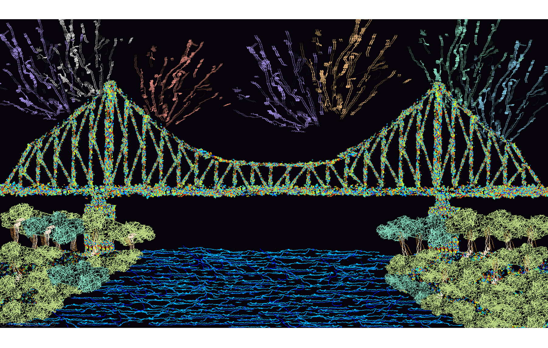
TrajectStory Bridge is a reconstruction of trajectories obtained from tracking individual proteins within a living cell over time, which maps out the route each molecule takes. Trajectories are assembled to depict the Story Bridge in Brisbane and projected onto a dark background, highlighting the intricacies of the molecular routes.
This artwork was constructed from several super-resolution images obtained from tracking individual proteins over time in live cells. This technique, called single-molecule imaging, allows us to understand how nanoscale dynamics and organisation of proteins can regulate biological processes. In this image, a tight junction protein, ZO-1, was tracked along tight junctions in a model blood-brain barrier system.
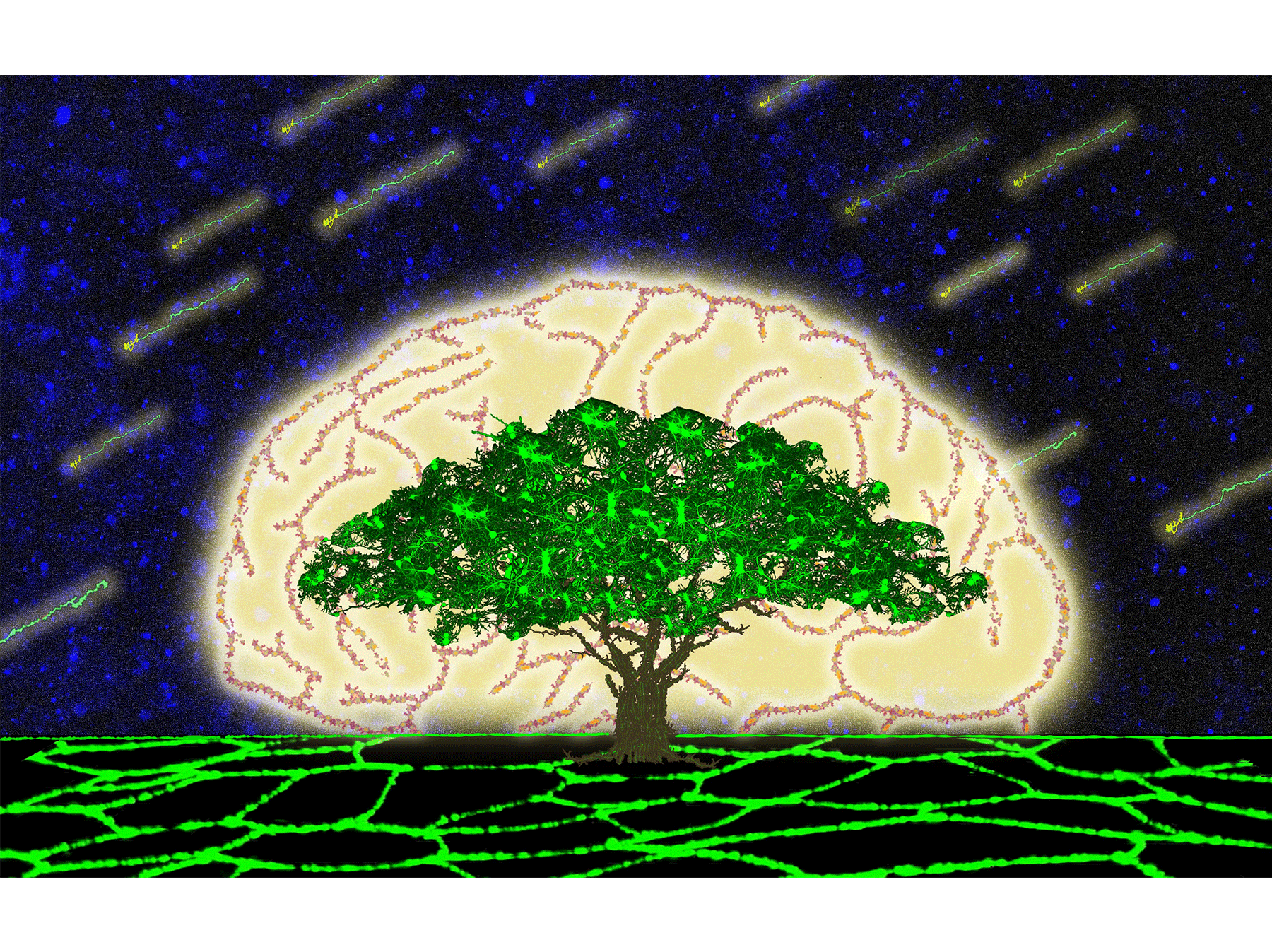
A neuronal tree flourishes above cell-cell junctions, depicting how the blood-brain barrier tightly controls the brain microenvironment to allow neurons to thrive.
Upon ultrasound insonification of microbubbles, a model drug (red) is delivered into cells (green) within a monolayer and the microtubule network (magenta) is transiently disrupted.
This video came from a project where I was trying to visualise the process of drug delivery into a cell by oscillation of a microbubble induced by ultrasound waves. Ultrasound and microbubble treatment is an emerging modality for transient delivery of neurotherapeutics to the brain. However, how the drugs get through the blood-brain barrier in response to this treatment and the resulting cellular response has not been characterised. Directly observing these processes using our system coupled with high-resolution microscopy allows us to evaluate the direct cellular response to balance the efficacy and safety of ultrasound treatment.
Dr Jonathan Lee's current work combines molecular biology, biochemistry and quantitative imaging techniques to unravel the nanoscale changes in the spatiotemporal organisation of key synaptic proteins implicated in neurodegenerative diseases.
During his PhD, he developed extensive expertise in super-resolution single-molecule microscopy and various other advanced microscopy techniques and analyses. He also developed a custom-built imaging platform to apply ultrasound to live cells within a high-resolution microscope. Using this platform, he identified a spatiotemporally interconnected sequence of biological responses underlying the different fates of sonoporated cells.
"I often see proteins that look or behave differently in the pathological state, such as in models of Alzheimer’s disease. Sometimes, these cells, or subcellular structures resemble objects or things we are exposed to in our everyday lives, which gives me inspiration for my artworks. Once, I even saw a cell shaped like Australia, including the little Tasmania positioned accurately at the bottom."




.png)